Echocardiogram
Also known as an ‘echo’, this is also a non-invasive test. Rather than looking at the electrical conductivity,rhythms of the heart, it can evaulate the pumping action of the heart and look more at the structure of the heart and the valves. It uses sound waves that echo against the structure of the heart to build up a detailed picture; it is similar to an ultrasound scan used during pregnancy.
A lubricating jelly is put on the chest and then a probe (known as a recorder) is placed on the chest and moved about. This passes sound waves through the body and the probe picks up the echos reflected back from the various parts of the heart and shows this as a moving picture on the screen. Doctors can use this test to evaluate congenital heart problems, heart failure, valve disease and heart attacks.
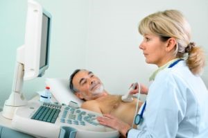
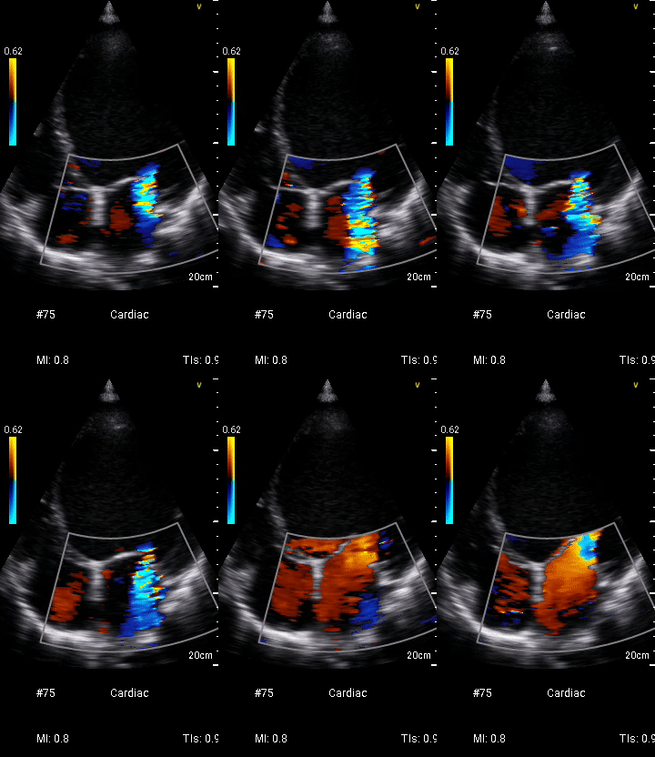
Echocardiography (ECHO) imaging.
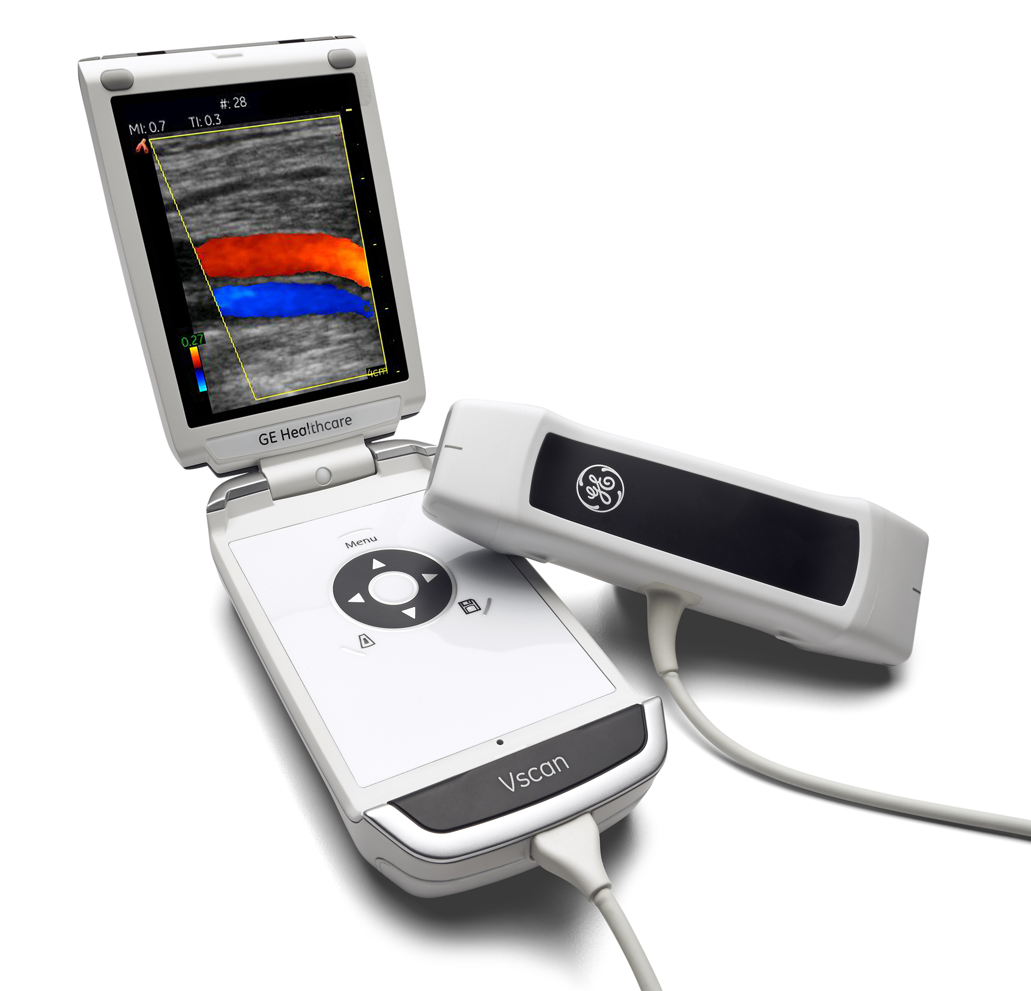
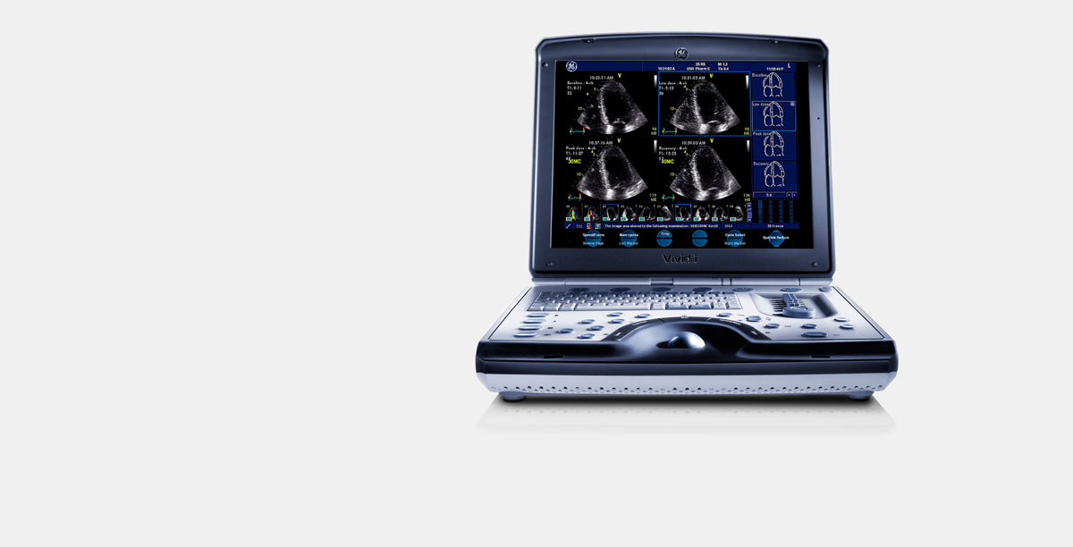
We use GE Healthcare scanning equipment for 2D imaging, colour flow and doppler assessment of heart muscle and valve function
